


Machine Learning Sleep EEG Measures Brain Age and Dementia Risk
By Irene Yeh
Sleep disturbances are emerging as early indicators for dementia. However, the broader architecture of sleep shows inconsistent associations between cognitive impairment and incident dementia. These broad sleep metrics don’t fully capture the complex and multidimensional nature of sleep physiology, according to Yue Leng, MBBS, Ph.D., associate professor of psychiatry at the University of California, San Francisco School of Medicine. Fortunately, sleep electroencephalography (EEG) can capture detailed patterns of brain activity that directly reflect underlying neural processes and their functions, detecting dementia before symptoms appear while also providing insight into how cognitive abilities change with aging.
Changes in EEG microstructure usually follow a predictable pattern as people age, but some individuals may show faster decline in these patterns. Although previous studies have shown a correlation between cognitive impairment and multiple sleep EEG patterns (e.g. spectral power, sleep depth, and spindle-slow oscillation coupling), huge amounts of EEG patterns are challenging to summarize and interpret.
To overcome this barrier, Leng and her team developed a sleep EEG-based brain age using a first-of-its-kind interpretable machine learning approach that integrates multiple age-dependent EEG microstructures into a single age-like number. They then computed the brain age index (BAI)—the difference between brain age and chronological age—with sleep EEG microstructures. They investigated the connection between BAI and dementia risk across five cohorts and whether key dementia risk factors influenced this association. Their findings were published in JAMA Network Open (DOI: 10.1001/jamanetworkopen.2026.1521).
The Relationship Between BAI and Incident Dementia
The study included five cohorts from the Multi-Ethnic Study of Atherosclerosis with 956 females and 846 males; the Atherosclerosis Risk in Communities study with 918 females and 878 males; the Framingham Heart Study–Offspring Study with 318 females and 299 males; the Osteoporotic Fractures in Men Study with 2,639 males; and the Study of Osteoporotic Fractures with 251 females. The team notes that the majority of the participants across all cohorts were cognitively normal at the time of sleep assessment.
While the large participant pool meant a diverse population, it also meant the pool composed of different population characteristics, data collection methods, dementia assessment, and follow-up durations, which may introduce heterogeneity and cause potential bias when combining results from all studies.
The results found that 1,082 participants had incident dementia. Across the cohorts, each 10-year increase in BAI was linked to a 39% higher risk of incident dementia. Even after accounting for factors such as age, sex, race and ethnicity, sleep medication use, physical activity level, education, smoking status, and BMI, the risk remained the same at 39%. The team also adjusted for cognitive scores, diabetes, high blood pressure, heart attacks, stroke, depression, and Apnea-Hypopnea Index score at the time of sleep assessment, and the association barely decreased. These results may suggest that the link between BAI and subsequent dementia is separate from cognitive status and many comorbidities.
Brain Age and Dementia Risk
Previous studies conducted by the team confirmed sleep-based BAI in clinical settings. The present study’s findings show, for the first time, that BAI’s association to future dementia risk applies to people outside clinical communities. Rather than predicting dementia, BAI is trained on large EEG datasets across the lifespan using a person’s actual age as the reference. This allows the use of a much larger and more diverse data pool than would be available for dementia-focused models. As such, the results reinforce BAI’s ability as a useful marker for accelerated brain aging.
Other biological mechanisms may explain why higher BAI correlates with increased risk of dementia, such as high levels of tau proteins in cerebrospinal fluid and greater amyloid buildup being associated with changes in sleep-related brain activity. Leng and her team found that adjusting for APOE genotype had little to do with the relationship between BAI and dementia risk, indicating that BAI is not just reflecting genetic risk for Alzheimer’s disease.
A limitation of the study is the inclusion of only death as a competing risk for dementia. Other life events, such as major surgeries, psychiatric illnesses, or acute medical conditions may affect follow-up or dementia ascertainment and warrant consideration as additional competing risks.
Additionally, because the study is observational, it cannot infer a causal relationship between BAI and dementia. As a composite measure, BAI is not a plausible therapeutic target and should be viewed as a prognostic marker for future dementia risk. If an individual shows an older BAI than their age would indicate, it is recommended to conduct an examination of their specific EEG microstructural components to determine what is driving this deviation, interpret their neurophysiological significance, and assess whether these underlying processes may represent viable therapeutic targets.
The reliance on EEG microstructural features for BAI estimation may also limit broader applicability. The team writes that future research should use wearable devices for validation to ensure broader generalizability and support clinical implementation. BAI not only provided insights to neurophysiological signals that reflect future dementia risk or resilience, it also may help with identifying people who need closer cognitive monitoring, identify high-risk patients, and better inform clinical decision-making.
About the Author: Irene Yeh
Also on Integrative Practitioner

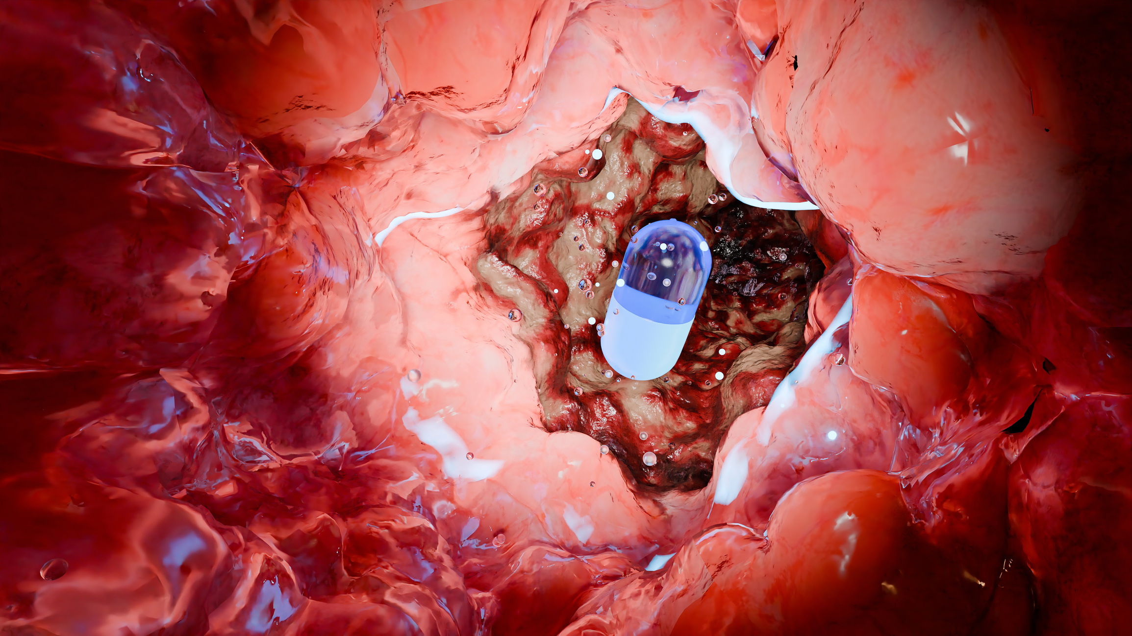
Vibrating Pill Could Both Treat Eating Disorders and Predict Relapse
By Deborah Borfitz Digestive sensations profoundly affect mental health through the bidirectional gut-brain axis, and in the case of anorexia nervosa (AN)...

Fluorescent Nanosensor Offers Rapid Way to Measure Gut Health Biomarker
By Integrative Practitioner Staff Researchers from the SMART Innovation Centre, the National Institute of Education (NIE), and Nanyang Technological University,...

Traditional Chinese Medicine May Help Relieve Long COVID Symptoms
By Irene Yeh The World Health Organization (WHO) defines long COVID as persistent symptoms of the COVID-19 virus that occur...

No Such Thing as “Moderate” with Alcohol Consumption
By Irene Yeh Alcohol consumption remains one of the leading causes of disease and death in the U.S., yet there...

New Alzheimer’s Blood Test Flags Symptom Onset
By Allison Proffitt Researchers from Washington University School of Medicine, St. Louis have showed that elevated levels of a set of circular RNAs (circRNAs) in the...