


Red Light Therapy May Help with Brain Recovery
By Irene Yeh
February 5, 2026 | Awareness about repetitive head acceleration events (RHAEs) has increased over the years, but treatment and prevention strategies remain limited. RHAE refers to repeated external collisions that rapidly move the head, either through direct contact to the skull or through impacts on the body that cause the head to move. Accelerated head events can result in poorer cognitive performance, abnormal brain activation patterns, decreased white matter integrity, and increased neuroinflammatory biomarkers. The chronic effects are less understood, even though it is estimated that between 125 and 440 head acceleration events occur for every one sport-related concussion in collegiate and professional football players, and individual players experience up to 77 head acceleration events in a single season (Journal of Neurotrauma, DOI: 10.1177/08977151251403554).
Current treatment only focuses on post-injury care, such as rest, symptom management, and exercise. Even with helmet improvements and rule changes to games, it is not enough to prevent RHAEs. Furthermore, current treatments do not address microscopic damage or toxic processes that unfold over time.
To find an effective care method, researchers at the University of Utah Health tested photobiomodulation (PBM), which shines near-infrared light through LEDs or lasers to specific brain areas. Also called red light therapy, this exposure activates mitochondrial responses that boost energy production in cells and improve blood flow, thus delivering more oxygen and nutrients to damaged tissues.
Testing It Out on Football Players
The researchers tested PBM on 26 football players from a National Collegiate Athletic Association (NCAA) Division I American university located in the Western U.S. The participants were evenly divided between an active or sham PBM group. The participants received a Neuro Gamma (v3) transcranial plus intranasal PBM device and given instructions on its use and maintenance. Treatments were self-administered under supervision of research staff in the training room or in a private space when traveling for away games. The devices were identical externally, but the sham devices were not emitting any red light. Treatments were 20 minutes long and administered three times a week over the course of 16 weeks.
A Significant Difference
Using correlational tractography, the research team identified significant differences in pre- and post-season changes in brain inflammation and damage to nerve fibers between the active PBM and sham PBM groups. The sham group showed widespread increases in restricted diffusion imaging (RDI) and quantitative anisotropy (QA)—brain injury markers. In contrast, the active group had mostly stable levels of these makers. These results suggest that PBM helps protect the brain from changes linked to RHAEs and may reduce the effects of past incidents, although long-term studies are needed to confirm lasting benefits.
The increases in RDI and QA in the sham PBM group match previous research that showed RHAEs can cause neuroinflammation and axonal damage, even within one sports season. Higher levels of RDI indicate more immune cells entering the brain, and elevated QA levels suggest the brain is trying to repair small structural damage in response to inflammation. Stable or lower levels of RDI and QA in the active PBM group indicate that red light therapy reduced inflammation and protected the brain from the damage that followed, including nerve cell injury or loss, reduced neuroplasticity, and long-term cognitive and mood issues.
Consistent use of red light therapy may also help with acute inflammatory effects of RHAEs within a sports season, and if it is applied across multiple seasons, potentially can reduce the cumulative effects of brain inflammation that contributes to long-term neurodegeneration. As previously mentioned, further studies are needed to confirm this.
One key finding is that the brain areas most influenced by PBM are the same areas known to be especially vulnerable to repeated head impacts, or the “cone of vulnerability.” These areas—the midbrain, brainstem, thalamus, basal ganglia, corpus callosum peripheral association tracts, and association tracts with frontal and temporal lobe terminations—experience the most physical stress during repeated head movements. The sham group showed the most inflammation and nerve fiber repair in these regions of the brain while the active group had smaller changes and decreases in injury markers, particularly in the parts of the back left side of the brain.
The study had a small sample size, and researchers were unable to report player positions without risking participant confidentiality. Recent studies have shown that player position can influence long-term effects of repeated head impacts, and this likely applies to RHAEs, as well. Additionally, the sample did not include a noncontact or limited contact sport group for comparison, which limited the researcher’s ability to determine if the observed neuroinflammation and microstructural changes can be attributed to RHAEs alone. However, the use of permutation testing, consistent results across multiple supplemental analyses, and the anatomical plausibility of the affected brain regions offer enough support to the findings.
About the Author: Irene Yeh
Also on Integrative Practitioner

Tracing Sauna’s Mechanism of Action for Health
By Allison Proffitt Researchers in Finland have sought to quantify how the practice of Finnish sauna bathing (FSB) contributes to...
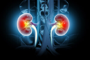
The Connection Between Oral and Kidney Health
By Irene Yeh Oral diseases have been linked to chronic kidney disease (CKD) due to inflammatory and microbial pathways that...

‘Humble’ AI Reveals When It is Uncertain in Diagnoses
By Irene Yeh Artificial intelligence (AI) models have assisted doctors with several clinical tasks, and they hold great promise in...
You Are What You Eat: Nutrition’s Impact on Mental Health
By Irene Yeh It is widely known that nutrition plays a crucial role in brain function, mood regulation, and overall...

AI is Here to Assist, Not Replace
By Irene Yeh As artificial intelligence (AI) continues to enter clinical spaces, there is an understandable concern around AI overtaking...