


Integrative assessments and approaches to osteoporosis
Photo Cred: Nino Liverani/Unsplash
By Kim Furtado, ND
Osteoporosis is considered a silent condition. Women are often diagnosed with osteopenia and feel helpless about managing bone loss. Many will take the recommendations, increasing weight-bearing exercises and implementing calcium and vitamin D supplements. However, several patients decline pharmaceutical interventions due to concerns about efficacy and side effects.
Integrative health practitioners can address osteoporosis in a holistic, comprehensive way. When I teach my patients about their bones, I describe two teams that are actively working, the osteoblasts and osteoclasts. Bone is a dynamic, not stagnant tissue. Osteoblasts are bone cells that form new bone, and osteoclasts break-down bone to release minerals into circulation. Both actions are always occurring in bone tissue.
A dual energy x-ray absorptiometry (DEXA) scan is used to measure bone loss. When a patient’s DEXA score is less than -2.4, she is diagnosed with osteoporosis. This score does not indicate that bone building is not occurring. It simply indicates that the rate of bone break-down is happening at a faster rate than bone-building, and that is quantified by the score in the scan. Through various factors including lifestyle, nutritional, hormonal, and environmental influences, the osteoclasts’ actions are more dominant than the osteoblasts’ actions. The most serious health concern related to osteoporosis is the increased risk of fracture, which can lead to chronic pain, long-term disability, and death.
My first step is to figure out what factors are helping the osteoclasts. The initial laboratory work-up includes salivary adrenal hormone testing, such as circadian cortisol, dehydroepiandrosterone (DHEA), estradiol, progesterone, and testosterone. Long-term chronic stress, as well as menopausal changes in hormonal levels, are known factors that accelerate bone loss.
Air pollution causes systemic as well as tissue-specific inflammatory changes and may be a factor in the pathogenesis of osteoporosis. Cadmium, fluoride, smoking, and lead are known contributing environmental factors for osteoporosis.
Since it is difficult to assess a client’s specific body burden in relationship to all known factors, I find provoked toxic element body burden testing can be useful. Thyroid function, iron, ferritin, and vitamin D levels are also beneficial tests.
When able, I will also encourage patients to monitor progress of treatment with at least quarterly urinary tests of pyrilinks-D, normalized to creatinine. Over time, we can watch for trends in the bone breakdown process.
Nutritional intervention should encourage high levels of mineral-rich foods, and reduce high amounts of animal protein, caffeine, soda, and alcohol, which are known disruptors of absorption or retention of minerals. The deeper dive includes understanding risks posed by acid-blocking medications, and improving digestion through bitters, mineral-infused vinegars, or digestive enzymes and betaine hydrochloric acid.
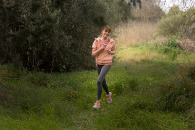
Each person’s appropriate nutritional and digestive functional medicine analysis is relevant to the osteoporosis work-up. Weight-bearing exercises is a great boon for the osteoblasts. It is also advisable to provide some added support such as complex partially hydrolyzed collagen and its associated proteins including bone morphogenetic proteins (BMPs).
For fracture prevention purposes, practitioners may focus on balance exercises. This may be obtained through general at-home exercises such as foot taps, standing marches and sits to stands. Time-honored practices such as yoga, or tai chi will also provide improved core strength and balance.
After an initial review of findings, I rely on the naturopathic principles to stimulate the body’s own ability to heal. By assessing individuals with labs to understand the factors that influence the osteoblasts and osteoclasts, treatment can be targeted to stimulate that patient’s innate healing process.
Case Study
Barbara is a 57-year-old female who presented with recent diagnosis of a compression fracture in the lumbar spine due to osteoporosis. After a recent recovery from an upper respiratory infection with cough, she had onset of severe back pain, which was subsequently diagnosed as fracture. A follow-up DEXA scan showed a -2.7 T-score of the lumbar spine. She was diagnosed with osteopenia at age 45, having a strong family history of osteoporosis, and an early menopause at age 45. She said she was told she was too young to take hormone replacement therapy, and her hormones were never subsequently tested despite seeing a gynecologist annually. She also noted a 26-year smoking history, which ended 10 years ago.
Her self-described diet intake was a “heart healthy” diet, consisting of whole grains, fruits, and vegetables with minimal animal protein. She reported “horrible” reactions to diesel fumes from a boat or bus, after exposure feeling progressively worse for two days. She described managing high cholesterol with a statin medication for several years, however recently made a change to use red yeast rice. Otherwise, she was an active, fit person whose exercise habits include Pilates, running, biking, swimming, and strength training. She also reported ongoing intake of calcium supplements since being first diagnosed with osteopenia.
Her initial treatment plan was geared to boost bone building and address the healing needed from the fracture. Her plan included digestive enzymes with ample amount of betaine hydrochloric acid, a comprehensive bone mineral formula in powder form for easy assimilation, and complex partially hydrolyzed collagen and its associated proteins including bone morphogenetic proteins (BMPs).
Upon her four-week follow-up, Barbara reported significant reduction of pain. Her follow-up x-ray showed her fracture was healing very well.
With our review of findings, her underlying hormonal imbalances were revealed, specifically with high morning cortisol, low estradiol, low DHEA, and elevated urinary pyrilinks-D, normalized to creatinine, creating an environment that favors bone loss. Moreover, her urine toxic elements showed lead levels four-times the reference range, and mercury was much higher than the reference range.
We continued to support adequate absorption and supply of minerals but began a two-month protocol for detoxification that included adequate nutrient, antioxidant support, liver supportive herbs and nutrients, and utilized DMSA as chelation agent. Some general adrenal support through 10 milligrams DHEA and eleuthrococcus senticus was also prescribed
After improving the patient’s liver function and detoxifying environmental toxins, the next focus is creating an environment for healthy bone building. I generally prefer to wait until inflammation levels are lowered through detoxification, and adrenals are nourished to re-test estradiol. If we can promote a woman’s ability to balance hormones with nutrition and adrenal support, then the bones can benefit without the risks of bioidentical hormone replacement therapy. At her nine-month check, I will re-visit and discuss with the patient what options are available for optimizing hormone levels.
In the big picture, I prefer to empower patients with osteoporosis to listen to known factors that affect their capacity to build and maintain their bones, and treat on an individual basis.
References
Apostu, D. (2020) Influence of Thyroid Pathology on Osteoporosis and Fracture Risk: A Review. Diagnostics. Retrieved from: https://www.mdpi.com/2075-4418/10/3/149
Chang, K. H., Chang, M. Y., Muo, C. H., Wu, T. N., Hwang, B. F., Chen, C. Y., Lin, T. H., and Kao, C. H. (2015). Exposure to air pollution increases the risk of osteoporosis: a nationwide longitudinal study. Medicine Retrieved from: https://insights.ovid.com/article/00005792-201505010-00014
Eman R. Youness, Nadia A. Mohammed and Fatma, A. (2012) Cadmium impact and osteoporosis: mechanism of action. Toxicology Mechanisms and Methods. Retrieved from: https://www.ncbi.nlm.nih.gov/pubmed/22708652
Lv, Y.. (2017) Cadmium Exposure and Osteoporosis: A Population-Based Study and Benchmark Dose Estimation in Southern China. Journal of Bone and Mineral Research. Retrieved from: https://asbmr.onlinelibrary.wiley.com/doi/full/10.1002/jbmr.3151
Lu, Y. H., Rosner, B., Chang, G., & Fishman, L. M. (2016). Twelve-Minute Daily Yoga Regimen Reverses Osteoporotic Bone Loss. Topics in geriatric rehabilitation. Retrieved from: https://insights.ovid.com/article/00013614-201604000-00003
Martini, C.N., Gabrielli, M., and Bonifacino, G. (2018) Lead enhancement of 3T3-L1 fibroblasts differentiation to adipocytes involves ERK, C/EBPβ and PPARγ activation. Molecular and Cellular Biochemistry. Retrieved from: https://link.springer.com/article/10.1007%2Fs11010-017-3093-y
About the Author
About the Author: CJ Weber
Also on Integrative Practitioner
Dental Danger: Signs of Sleep Breathing Disorders in children and Adults, Learn the Visual Clues
Non-Member Price: $30.00 Member Price: $18.00 – 40% Off for Members Only Add to Cart Presented by: Mark Breiner, DDS,...

Integrative and Comprehensive Approach to Inflammatory Bowel Disease
Non-Member Price: $30.00 Member Price: $18.00 – 40% Off for Members Only Add to Cart Presented by: Ronald Hoffman, MD,...

Measuring Biology vs. Symptomatology: The Fully Integrated Functional Wellness Program
Non-Member Price: $30.00 Member Price: $18.00 – 40% Off for Members Only Add to Cart Presented by: Dane Donohue, DC...

Change Your Genes, Change Your Life
Non-Member Price: $30.00 Member Price: $18.00 – 40% Off for Members Only Add to Cart Presented by: Kenneth Pelletier, PhD,...

Guided Imagery – Efficacious, Portable, Scalable, User-Friendly, Self-Administered
Non-Member Price: $30.00 Member Price: $18.00 – 40% Off for Members Only Add to Cart Presented by: Belleruth Naparstek, ACSW...