


Chiropractic approach to joint disease of the spine
Photo Cred: Freepik
By James Lehman
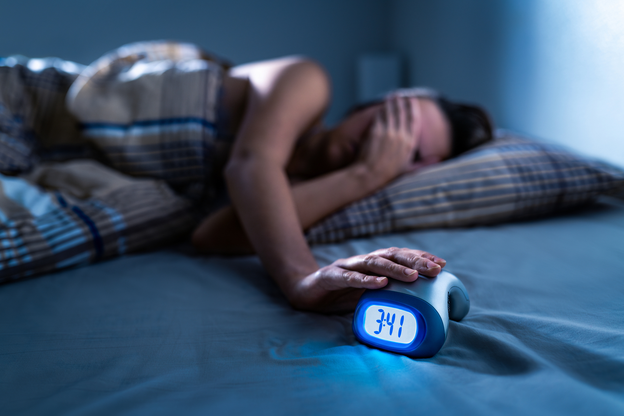
Any physician or healthcare provider that treats adults for painful spinal conditions must be familiar with degenerative joint disease of the spine. As stated by Joseph Pilates, “a man is as young as his spinal column.” As a chiropractic specialist, I concur with Pilates. A degenerating spine affects both function and quality of life.
Degenerative spinal joint disease, is commonly referred to as osteoarthritis, which is a disease of the spinal joints. It is the most common form of arthritis, affecting some 27 million American adults and usually follows trauma with injury to spinal muscles, tendons, ligaments, and joints.
When evaluating a patient with spinal pain or dysfunction, it is essential to perform a focused history of the present illness that reveals the location of the pain, mechanism of injury, onset, what makes the condition better and worse, and the description of the pain. If there is pain, is it localized to a specific level of the spine, or is it referred or radiating pain? Spinal pain should be rated for severity at its best and worst. I prefer the 11-point numerical rating scale, but there are a number of different pain scales available for your use to help determine if the condition is getting worse with more frequency. Finally, ask the patient what treatments were received and the outcomes.
Oftentimes, patients do not recall spinal injuries, including spinal strains and sprains that occurred years prior to their initial evaluation with you. It is human nature to forget painful events. You may need to further prod the patient and attempt to remind them of previous injuries to the spine with an explanation that describes how a spinal strain or sprain may affect the spinal anatomy and function.
Let me offer a classic example to make my point. The patient denies any previous injury that may have caused a spinal problem. Upon further investigation, you reveal that the patient had fractured one of their lower extremities as a young adult. While the patient may not realize how a fractured leg may cause spinal trauma and resultant spinal degenerative joint and disc disease, your examination may be provide convincing evidence.
When you observe a limping gait and postural evaluation reveals pelvic obliquity, I suggest you perform the Long Sit Test. This test will differentiate a functional short leg versus an anatomical short leg. If this patient demonstrates an anatomical short leg of the previously fractured lower extremity you should perform tape mensuration. This measurement illustrates the previously fractured lower extremity to be one inch shorter than the opposite one with our putative patient’s case. Now, you have the physical evidence that might depict the cause of the spinal degenerative changes. Although the fracture of the lower extremity did not cause injury to the spine, the long-term postural and gait abnormalities produced pelvic obliquity and repetitive spinal strain or sprain injuries and biomechanical joint dysfunctions with eventual degenerative changes in the anterior or intervertebral discs and posterior joints or facets.
Fortunately, with this patient it is possible to reduce the pelvic obliquity and improve the spinal joint function with placement of a half inch heel lift in the shoe of the short leg. Hopefully, this orthotic will alter the biomechanics and prove to be a successful intervention without the need for medications or surgery. This example illustrates the need to make the right diagnosis in order to provide the appropriate treatment.
While performing patient interviews, the clinician should be differentiating the list of potential diagnoses responsible for the patient’s complaint of spinal pain and/or dysfunction. Some of the most common spinal degeneration conditions are provided for your consideration:
- Post-traumatic spinal degeneration
- Posterior joint (zygapophyseal)
- Degenerative spondylolisthesis
- Spinal stenosis
To prevent negative outcomes when treating patients with spinal degeneration, clinicians must recognize neurological deficits caused by spinal stenosis. Patients may present with mild to moderate symptoms of clumsiness or inability to dress or undress because of the inability to manipulate buttons. The ability to walk may be limited by pain and/or weakness in the legs, loss of balance, and bowel or bladder dysfunction with the more severe symptoms.
Conservative care including spinal manipulation, acupuncture, massage, exercises, weight control, proper nutrition, and hydration are often effective with the above conditions providing there are no long tract signs indicating compression of the spinal cord, which may occur with spinal stenosis. Narrowing of the vertebral canal may compress the spinal cord and create an upper motor neuron lesion, which must be revealed during the patient history and neurological examination. The following neurological characteristics of an upper motor neuron lesion must be recognized in order to avoid permanent neurological deficits.
- Weakness without atrophy
- Hyper-reflexia
- Pathological reflexes (Babinski and/or Hoffmann’s signs)
- Clonus
- Spastic paralysis
Unlike spinal degeneration with subsequent biomechanical dysfunction and localized joint pain, spinal stenosis with spinal cord compression usually requires referral to a neurosurgeon for consultation. Spinal manipulation may be either an absolute or relative contraindication when patients present with moderate to severe symptomology. These patients may benefit more from surgical intervention rather than conservative care.
References
Campbell, T., Ghaedi, B., Ghogomu, T., and Welch, V. (2018) Shoe lifts for leg length discrepancy in Adults with common painful musculoskeletal conditions: A systematic review of the literature. Archives of Physical Medicine and Rehabilitation.
Snyder, D., Doggett, D., and Turkelson, C. (2004) Treatment of Degenerative Spinal Stenosis. American Family Physician.
Van Jign, J. (2002) Babinski Sign. Practical Neurology.
Editor’s note: Photo couresty of Freepik.
About the Author: CJ Weber
Also on Integrative Practitioner
Dental Danger: Signs of Sleep Breathing Disorders in children and Adults, Learn the Visual Clues
Non-Member Price: $30.00 Member Price: $18.00 – 40% Off for Members Only Add to Cart Presented by: Mark Breiner, DDS,...

Integrative and Comprehensive Approach to Inflammatory Bowel Disease
Non-Member Price: $30.00 Member Price: $18.00 – 40% Off for Members Only Add to Cart Presented by: Ronald Hoffman, MD,...

Measuring Biology vs. Symptomatology: The Fully Integrated Functional Wellness Program
Non-Member Price: $30.00 Member Price: $18.00 – 40% Off for Members Only Add to Cart Presented by: Dane Donohue, DC...

Change Your Genes, Change Your Life
Non-Member Price: $30.00 Member Price: $18.00 – 40% Off for Members Only Add to Cart Presented by: Kenneth Pelletier, PhD,...

Guided Imagery – Efficacious, Portable, Scalable, User-Friendly, Self-Administered
Non-Member Price: $30.00 Member Price: $18.00 – 40% Off for Members Only Add to Cart Presented by: Belleruth Naparstek, ACSW...